

In collaboration with the Jaffe Lab at Scripps, Smith Lab Postdoctoral Scholar, Or Ben-Zvi, has developed a cutting-edge microscope to study coral photosynthesis and health.
The diver-operated microscope — called the Benthic Underwater Microscope imaging PAM, or BUMP — incorporates pulse amplitude modulated (PAM) light techniques to offer an unprecedented look at coral photosynthesis on micro-scales.
In a new study, Ben-Zvi et al. describe how the BUMP imaging system makes it possible to study the health and physiology of coral reefs in their natural habitat, advancing longstanding efforts to uncover precisely why corals bleach.
Read more at the following sites:

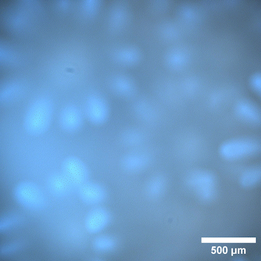
An image of Montipora coral polyps taken by the BUMP microscope. Credit: Or Ben-Zvi
A white illumination video of the coral Porites taken in-situ in Maui, Hawaii, under ambient light. In the video, the top left polyp is reacting to a particle that passes through by contracting its tentacles. Credit: Or Ben-Zvi